Homepage

If you have landed on this page it is highly likely that you’re an A-Level student, or someone of a similar level, and you’re interested in taking your understanding of the neurological disorder ‘Amnesia’ to the next level.

If you feel that your current understanding of amnesia isn’t of an A-Level standard, or you feel you might need a quick refresher on the fundamental understanding of amnesia (particularly retrograde and anterograde), then the below link may help you get up to speed before continuing this article.

https://gcsepsychology.com/brain-damage-retrograde-anterograde-amnesia/

To better help apply the information in this wiki to A-Level studies, two different amnesia disorders will be referred to for examples on each of the key pages. These are: Wernicke-Korsakoff Syndrome (WKS) and Transient Global Amnesia (TGA). Other disorders may be referenced throughout but the three mentioned above will be the focus.

To fully understand amnesia, the processing of memories must first be understood. Briefly, short-term memory (STM) is defined as information held within conscious awareness, and which is currently receiving attention; it is of course contrasted to long-term memory (LTM) which is held in permanent storage available for retrieval at some time in the future (Groome, 2013, p. 137). For example, when revising for an exam the information the student is currently studying would be present in their short-term memory but the information they learnt in class at the start of the year that they haven’t revised yet would likely be in their long-term memory stores, if not forgotten.

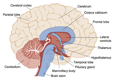
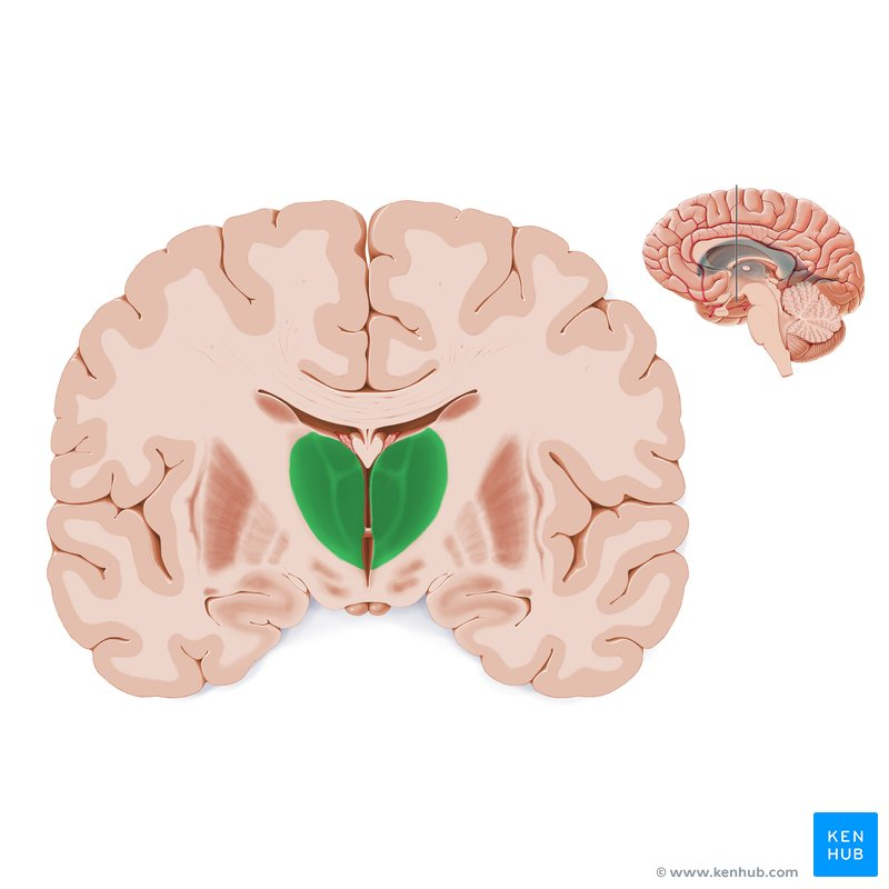
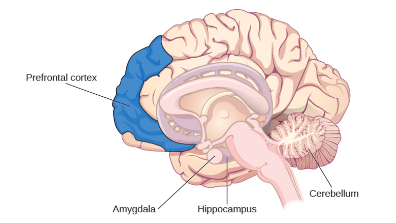
Many researchers believe that the entire brain is involved with memory, in some way or another (Lashley, 1950). However, some key areas have been identified (shown below in Figure 1.)  and include the amygdala, hippocampus, cerebellum, the prefrontal cortex and the synapses linking them all (Mayford, Siegelbaum, & Kandel, 2012).

Figure 1. Identifying key structures involved in memory processing

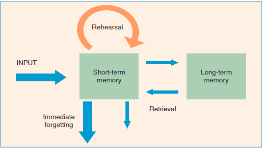
Forming memories involves various steps and stages (Atkinson & Shiffrin, 1968). To begin with the brain receives sensory information from several sources in different formats, such as smell, taste, sound, vision and touch. At this point the information that has the individuals focus will be rehearsed in the STM store and ‘encoded’ (transformed into a format the brain can retain). Following this, the information is then ‘committed to memory’ and stored in the LTM, readily available to be retrieved when needed. The information not rehearsed or not regularly retrieved will eventually be forgotten. Figure 2. Below illustrates this process.

Figure 2. Illustrating Atkinson & Shiffrin (1968) theory on memory processing

Amnesia is essentially the instances where memory, or the process of forming memories, fails. The processes of forming, maintaining and recalling memories is extremely complex and as mentioned above involves numerous brain structures, if not the brain in its entirety. This can lead to numerous points of failure and thus numerous reasons for memory to fail; leading to amnesia.

In terms of understanding amnesia and memory, the convoluted and complex nature of the memory process can make it difficult to pinpoint which areas are responsible for which functions, this problem is also relevant when looking at the symptoms of amnesia which quite often include several deficits outside those of memory. These are referred to as ‘comorbid’ symptoms, diseases or conditions that occur alongside another disorder, and can make it difficult to differentiate the issues that are a direct result of amnesia and those that are not. Examples of those that appear alongside amnesia would be perceptual deficits and confabulation; this is often associated with WKS and will be further described in the ‘Signs/Symptoms’ section below.

However, as research has developed, and the mapping of the brain and its functions has developed researchers are becoming able to pinpoint the structure’s role within memory, such as the amygdala facilitating the encoding of memories to a more significant degree when the event is emotionally arousing (Josselyn, 2010). This means that when an individual suddenly fails to respond to stimuli that normally would have induced a fear response; a neurologist can narrow down their investigation and begin imaging of the amygdala with the expectation of some sort of abnormality (e.g. a lesion or tumour) affecting it’s functioning.

Once more, if further understanding of Memory and some of the key structures involved (such as the ones mentioned above) is required the below link may be of some use.

https://courses.lumenlearning.com/wsu-sandbox/chapter/parts-of-the-brain-involved-with-memory/

Signs/Symptoms

Wernicke-Korsakoff’s syndrome (WKS) has been defined as “an abnormal mental state in which memory and learning are affected out of all proportion to other cognitive functions in an otherwise alert and responsive patient” (Victor, Adams, & Collins, 1971). This refers to the dramatic deficits in memory, and the related functions, that WKS patients display whilst maintaining proficient use of most of their other cognitive faculties. Many of the questions surrounding WKS, from as long as 50 years ago, remain. This could be because WKS is a syndrome resultant of numerous symptoms rather than a distinctive condition in it’s own right. This would explain the variety of symptoms that WKS patients may display, as well as the number of brain structures that could be involved. However, there are still some signs and symptoms that seem to have become rather typical of WKS.

Typically, WKS patients are poor at paired-associate and this can be indicative of the anterograde amnesia that is often present in WKS cases (Parkin, Dunn, Lee, Ohara, & Nussbaum, 1993). Paired-Associate learning is where individual is given paired words to learn and is later given on of the words and asked to recall the word it was paired with. An example of a Paired-Associate Learning (Free Recall) task is linked below.

WKS patients often confabulate (Kopelman, 1987) and it occurs when an individual’s memory is so poor that they make up facts to fill in the gaps, genuinely believing what they say as truth (Matthews & McClelland, 2010). The events the individual will describe are often reasonable, they just may not have happened to the individual themselves or just didn’t happen when they thought they did. An example would be a student simply detailing, with absolute sincerity, what lessons they had in school yesterday, but it was the middle of summer and they hadn’t been to school in the last six weeks. Below is a video link that could be useful in further understanding the concept of confabulation and how to manage situations that involve an individual who may be confabulating, as well as other disorders in which it may occur.

TGA was first described by Fisher and Adams (1964), who suggested that it was a ‘…sudden of an anterograde amnesia and confusion that resolved within a few hours’.

This understanding has since been developed to suggest that TGA is often characterised by a sudden onset of both an anterograde and retrograde amnesia and can in fact last up to 24 hours (Portaro, et al., 2018). Whilst it is often associated with an impairment of executive function and recognition during the episode, there tends to be no other neurological deficits or lasting effects following the episode (Lin , Chen, & Fuh, 2014) (Jaffe & Bender, 1966). TGA patients are also more likely to exhibit irritable and anxious behaviour (Serafetinides, 1994).

Causes

It’s commonly believed, and portrayed in the media, that amnesia is nearly exclusively caused by physical damage to the structures of the brain. It is also likely that this is the main cause taught at GCSE level psychology, if not the only discussed cause.

While brain damage is arguably the most common of causes, such as in cases of WKS, however it’s not the only cause. In fact, one of the criteria to accurately diagnose TGA is the absence of evidence suggesting head injury/trauma (Hodges & Warlow, 1990). Where damage to brain structures is the cause however, it need not be a physical trauma, resulting from a car crash for example, but damage to structures as a result of a virus or prolonged substance abuse.

The currently accepted theory of WKS revolves around chronic alcoholism, paired with a poor dietary regime, leading to a thiamine (Vitamin B) deficiency. The poor dietary regime can cause Wernicke’s Encephalopathy (WE) which is an acute neuro-psychiatric condition caused by an inadequate supply of thiamine (vitamin B1) to the brain (Thomson, Guerrini, & Marshall, 2012). If left undiagnosed or not properly treated, WE can develop into WKS which is much more severe and is detailed in the previous ‘Signs/Symptoms’ section above.

Thiamine is essential in the metabolism of glucose (the process is shown below in Figure 3) in the brain; the deficiency causes a loss, and/or shrinkage, of the neurons (Kalat, 2016). It has been noted the in WKS patients this loss/shrinkage of neurons often seems to be comprised of cholinergic neurons where the brain damage occurs in the basal forebrain (Nardone, et al., 2010).

Figure 3. The role of thiamine in metabolism of glucose

However, the damage to the basal forebrain doesn’t explain the persistent amnesia in cases of WKS; there must be damage to other structures/areas. Malamud & Skillicorn (1956) suggested that it was lesions to the mammillary bodies (Figure 4) causing the amnesia, whereas Victor et al (1989) posed that it was in fact the Dorsomedial Thalamic Nuclei (DMTN) (Figure 5). More recent research has found that mammillary bodies or DMTN alone were insufficient to cause the amnesia associated with WKS; only if both had suffered damage did the amnesia occur (Zuccoli, et al., 2009).

Similar research has found that 80% of patients had evidence of symmetrical lesions to the medial thalami and periventricular region of the third ventricle (Zuccoli, et al., 2009). Figure. 6 below identifies some of these brain structures (A), as well as how they differ in ‘alcoholic’ and ‘non-alcoholic’ participants (B).

Figure 6. Identifying and comparing brain structure of ‘Alcoholic’ and ‘Non-Alcoholic’ WKS patients (Zuccoli et al, 2009)

It has been argued that issues with this explanation of WKS are that whilst poor diet and chronic alcohol abuse can quite reasonably contribute to a thiamine deficiency, it doesn’t explain the absence of WKS cases in famine situations where thiamine deficiencies are prevalent. Thomson, Guerrini & Marshall countered  (2012) thiamine deficiency leads to a reduction in the activity of thiamine-dependent enzymes, and in turn to alterations in mitochondrial activity, impairment of cell metabolism, decreased energy status and eventually selective neuronal death. The damage caused by the combination of thiamine deficiency and alcohol metabolism probably interferes with adequate thiamine transport at a number of sites in the body, including the blood–brain barrier, as well as causing damage to the inactive thiamine-dependent enzymes which then require higher concentrations of thiamine to work normally.; however, the issue of why non-alcoholics are able to develop WKS remains.

For other disorders different individuals can be at a greater risk to develop them, for example key risk factors of TGA have been identified and include a history of migraines, ischemic heart disease (Also known as coronary heart disease, when insufficient oxygen reaches the heart muscle), carotid atheromasia (The presence of a atheroma blocking the carotid artery) and psychophysical stress (The effect of psychological stress on the physical body) (Jang, Park, & Hong, 2014) (Agosti, Akkawi, & Borroni, 2006).

TGA episodes are most likely to occur females and in the 7th decade of life (with a mean age of 61-67.3 years), supposedly due to the fact risk factors and naturally associated pathologies have a higher rate of incidence (Arena, Brown, & Mandrekar, 2017).

Whilst the above risk factors have been suggested to increase the likelihood of a TGA episode, it has also been found that ‘close’ and ‘remote’ precipitating events can be triggers leading to an episode. Close precipitating events for TGA include emotional stress, physical effort/exertion (this includes sexual activity and orgasms (Lane, 1997)) and water contact/extreme temperature change; whereas remote precipitating events would include anxiety (particularly a work/home conflict), general health problems and even financial stressors (Erkelens & Snoek, 2010).

Research has also indicated to some specific biological causes of TGA. Such as a stress-induced catecholamine release may lead to hypoxia/ischaemia, or neurotransmitters involved in this release may affect the formation of memories (Finsterer & Stöllberger, 2017). Alternatively, Spiegel, McCroskey & Deyerle (2016) found that severe emotional stress may contribute to the destabilisation of the CA1 sector of the Hippocampus via a massive glutamate release; leading to a disruption in the formation of memories.

On the other hand, TGA episodes may also be considered a psychological defensive mechanism, in cases of psychological disorders such as PTSD and dissociative disorders, associated to a phobic personality trait (Portaro, et al., 2018). Furthermore, psychological stressors, a psychogenic cause, appeared to disturb the affective learning circuit between the amygdale, hippocampus, striatum and the prefrontal cortex. As well as affecting the inhibiting effects on the amygdala; disrupting the memory formation process (Spiegel, McCroskey, & Deyerle, 2016). It is important to consider both biological and psychological causes for the TGA as they are both prominent factors in cases; 90% of examined TGA episodes involved physical or psychological precipitating factors (Quinette, et al., 2006).

Diagnosis/Prognosis

WE is reversible if treated with a timely and adequate dose of parenteral thiamine. If it is undiagnosed, or treated with inadequate dosages of thiamine, it is likely to proceed to the chronic state, Wernicke-Korsakoff’s Syndrome (‘inadequate’ dosages are explained below in the treatments section). The challenge with WKS is to prevent the patient developing the syndrome in the first place, but if this fails, to manage WKS so as to improve brain function and to aid the patient to adapt to their cognitive impairment, suggesting that the cognitive deficits are irreversible in cases that have progressed too far; how far is too far remains unclear. The general method of diagnosis is the use of Magnetic Resonance Imaging (MRI) to identify brain damage detailed in the ‘Causes’ section should patients be showing an anterograde amnesia, as well as possessive a history of poor nutrition and alcoholism.

As mentioned, sometimes patients have already sustained irreversible brain damage at the time of presentation (Thomson, Guerrini, & Marshall, 2012). The accumulated damage, from thiamine deficiency and alcohol misuse, is likely to render the use of oral thiamine therapeutically inadequate since the body is unable to produce high enough concentrations of thiamine in the blood to traverse the blood–brain barrier.

Hodges & Warlow (1990) identified very clear criteria that must be met for an accurate diagnosis of TGA. These are listed below in Figure 7.

Figure 7. TGA diagnosis criteria (Hodges and Warlow, 1990)

The above criteria are critical because other disorders, such as Transient Epileptic Amnesia (TEA) and Transient Ischemic Attack (TIA) may involve similar episodes but whilst similar they are indeed definitively different. Furthermore, the requirement for strict diagnostics is further emphasised by the fact that TGA does not typically show acute changes when using brain imagining techniques, apart from T2-hyperintense punctate lesions in lateral hippocampal regions (Faust & Nemes, 2010). Thus, these techniques cannot be relied upon to aid a diagnosis as is the case with other disorders of amnesia.

The good news when examining TGA is that it is believed to be relatively benign leaving no residual damage following episodes and as earlier mentioned, without long-term conditions/deficits resulting from an episode (Portaro, et al., 2018). It is also worth noting that individuals do not often experience repeat episodes.

Treatments

The biggest issue when treating amnesia is that there is no remediation of lost memories; the memories lost, and those not fully formed, due to an amnesic disorder are often permanently lost (Squire, 2006). In some instances, the forgetting of amnesia is a result of loss of access/failures in retrieval of information (Macleod & Macrae, 2001). When the issue lies within access and retrieval, ‘reconsolidation’ and ‘reconciliation’ may lead to patients fully recovering memories that were once unobtainable (Power, Berlau, McGaugh, & Steward, 2006). Thus, treatment in these cases must be centred around addressing the disorder causing persistent amnesia; as oppose to trying to recover memories lost due to the disorder and providing patients with ongoing support.

The challenge with WKS is to prevent the patient developing the syndrome but if this fails, to manage WKS to improve brain function and to aid the patient in adapting to their cognitive impairment, suggesting that the cognitive deficits are irreversible in cases that have progressed too far; how far is too far remains unclear.

The straightforward treatment for WE and WKS is to simply supplement the diet with thiamine; administered orally. However, as previously mentioned it is often the case that oral treatment is inadequate in cases of WKS (Thomson, Guerrini, & Marshall, 2012). This could be for several reasons such as insufficient quantities; ingested nutrients are often lost to vomiting or diarrhoea caused by poor diet as well as the fact that metabolism of alcohol increases the body’s requirements of thiamine. Another reason could be impaired absorption, alcohol-related liver damage impairs the phosphorylation of thiamine (see Figure 1) which is vital for the active transport in the absorption of thiamine for cell metabolism (Thomson & Marshall, The Treatment of Patients at Risk of Wernicke Encephalopathy in the Community., 2006). Finally, this could be due to thiamine transport issues, prolonged damage by the above causes could affect transport, meaning thiamine concentrations high enough to traverse the blood-brain barrier cannot be produced (Thomson, Guerrini, & Marshall, Wernicke’s Encephalopathy: Role of Thiamine, 2009). These factors mean that effective thiamine supplementation must be administered via parenteral routes; not via the digestive tract i.e. intravenously.

Beyond the supplementation of thiamine, it is believed that treatment packages should be designed to maintain abstinence of alcohol in a supported housing/rehabilitation environment. Ongoing follow-ups are key to monitoring cognitive status as well as psychiatric and functional wellbeing (Leenane, 1986). A cognitive behavioural approach has shown to be useful in develop coping strategies for day to day routines, things such as writing down information, keeping a diary and breaking tasks down into smaller steps (distributive practice).

As stated, TGA tends to leave no lasting effects meaning that direct treatment of the neuropsychological effects would be largely unnecessary. Though it is advised that individuals document the events leading up to the episode, so they may address any underlying issues or take preventative measures. However, that’s not to say that no treatment takes place. The sudden nature of an episode can be quite distressing for the individual, especially as the cause/trigger may not be totally obvious at first glance, so some level of counselling and understanding may need to take place for the individual to fully recover.

Useful Contacts and Further Information

Below will be some direct links for further information relating to the study of amnesia, and related neuropsychological topics, as well as some sites that will be able to provide support and assistance with cases of amnesia. For further academic information and studies, see the ‘References’.

https://www.nhs.uk/conditions/memory-loss-amnesia/
– The NHS page for amnesia information, assistance, advice and preliminary diagnosis

https://www.psychologytoday.com/gb/counselling/dissociative-disorders/eng/london

-Link to professional counselling contacts, specifically for amnesia/dissociative disorders

https://www.butterflies.org.uk
– Getting support for cases of Dementia

https://www.headway.org.uk/about-brain-injury/individuals/effects-of-brain-injury/memory-problems/
– Dealing with the effects of brain injuries

https://m.youtube.com/watch?v=2fXpdasTNTE


– A useful summary video for Transient Global Amnesia

References


Agosti, C., Akkawi, N. M., & Borroni, B. (2006). Recurrency in Transient Global Amnesia: A Retrospective Study. European Journal of Neuropsychology, 13, 986-989.


Arena, J. E., Brown, R. D., & Mandrekar, J. (2017). Long-Term Outcome in Patients with Transient Global Amnesia: A Population-Based Study. Mayo Clinic Proceedings, 92, 399-405.
Atkinson, R. C., & Shiffrin, R. M. (1968). Human Memory: A Proposed System and its Controlled Processes. In K. W. Spence, & J. T. Spence, The Psychology of Leanring and Motivation (2nd ed., pp. 89-195). New York: Academic Press.


Erkelens, C. D., & Snoek, J. W. (2010). What Doctors Should Not Forget About Transient Global Amnesia. European Journal of General Practice(16), 182-185.


Faust, J. S., & Nemes, A. (2010). Transient Global Amnesia: Emergency Department Evaluation and Management. Emergency Medicine Practice(18), 205-214.


Finsterer, J., & Stöllberger, C. (2017). Transient Global Amnesia: The Cerebral Takotsubo? Journal of the Neurological Sciences, 196-197.


Fisher, C. M., & Adams, R. D. (1964). Transient Global Amnesia. Acta Neurologica Scandinavica. Supplememtum(9), 1-83.
Groome, D. (2013). An Introduction to Cognitive Psychology: Processes and Disorders (3rd ed.). Hove, East Sussex: Psychology Press.


Hodges, J. R., & Warlow, C. P. (1990). Syndromes of Transient Amnesia: Towards a Classification. A Study of 153 Cases. Journal of Neurology, Neurosurgery and Psychiatry, 53, 834-843.
Jaffe, R., & Bender, M. B. (1966). E.E.G Studies in the Syndrome of Isolated Episodes of Confusion with Amnesia: “Transient Global Amnesia”. Journal of Neurology, Neurosurgery and Psychiatry, 29, 472-474.


Jang, J. E., Park, S. Y., & Hong, J. H. (2014). Different Risk Factor Profiles Between Transient Global Amnesia and Transient Ischemic Attack: A Large Case-Control Study. European Journal of Neurology(71), 19-24.


Josselyn, S. A. (2010). Continuing the Search for the Engram: Examining the Mechanism of Fear Memories. Journal of Psychiatry & Neuroscience, 35(4), 221-228.
Kalat, J. W. (2016). Biological Psychology (12th ed.). Boston, MA: Cengage Learning.


Kopelman, M. D. (1987). Two Types of Confabulation. Journal of Neurology, Neurosurgery and Psychiatry, 50(11), 1482-1487.
Lane, R. J. (1997). Recurrent Coital Amnesia. Journal of Neurology, Neurosurgery and Psychiatry, 63, 260.
Lashley, K. S. (1950). In Search of the Engram. In Society for Experimental Biology, Physiological Mechanisms in Animal Behaviour (Society’s Symposium IV ed., pp. 454-482). Oxford, England: Academic Press.


Leenane, K. J. (1986). Management of Moderate to Severe Alcohol-Related Brain Damage (Korsakoff’s Syndrome). Medical Journal of Australia, 145, 136-143.


Lin , K. H., Chen, Y. T., & Fuh, J. L. (2014). Migraine is Associated with a Higher Risk of Transient Global Amnesia: A Nationwide Cohort Study. European Journal of Neurology, 21, 718-724.


Macleod, M. D., & Macrae, C. N. (2001). Gone But Not Forgotten: The Transient Nature of Retrieval-Induced Forgetting. Psychological Science, 12, 148-152.
Malamud, N., & Skillicorn, S. A. (1956). Relationship Between the Wernicke and the Korsakoff Syndrome: A Clinicopathologic Study of Seventy Cases. AMA Archives of Neurology and Psychiatry, 76(6), 585-596.


Matthews, P. M., & McClelland, J. L. (2010). The Memory Process: Neuroscientific and Humanistic Perspectives. Cambridge, MA: MIT Press.


Mayford, M., Siegelbaum, S. A., & Kandel, E. R. (2012). Synapses and Memory Storage. Cold Spring Harbor Perspectives in Biology, 4.


Nardone, R., Venturi, A., Golaszewski, S., Caleri, F., Tezzon, F., & Ladurner, G. (2010). MR Atypical Wernicke Encephalopathy Showing Extensive Brain Stem and Diencephalic Involvement. Journal of Neuroimaging, 20(2), 204-207.


Parkin, A. J., Dunn, J. C., Lee, C., Ohara, P. F., & Nussbaum, L. (1993). Neuropsychological Sequelae of Wernicke′s Encephalopathy in a 20-Year-Old Woman: Selective Impairment of a Frontal Memory System. Brain and Cognition, 21(1), 1-19.
Portaro, S., Naro, A., Cimino, V., Maresca, G., Corallo, F., Morabito, R., & Calabro, R. S. (2018). Risk Factors of Transient Global Amnesia: Three Case Reports. Medicine, 97(41).


Power, A. E., Berlau, D. J., McGaugh, J. I., & Steward, O. (2006). Anisomycin Infused Into the Hippocampus Fails to Block ‘Reconsolidation’ but Impairs Extinction: The Role of Re-Exposure Duration. Learning and Memory, 13, 27-34.


Quinette, P., Guillery-Girard, B., Dayan, J., de la Sayette, V., Marquis, S., Viader, F., . . . Eustache, F. (2006). What Does Transient Global Amnesia Really Mean? Review of the Literature and Thorough Study of 142 Cases. Brain: A Journal of Neurology, 129(7), 1640-1658.


Serafetinides, E. A. (1994). Transeient Epileptic Amnesia: A Clinical Update and a Reformulation. Journal of Neurology, Neurosurgery and Psychiatry, 57, 1549.


Spiegel, D. R., McCroskey, A. L., & Deyerle, B. A. (2016). A Case of Transient Global Amnesia: A Review and How it May Shed Further Insight Into the Neurobiology of Delusions. Innovations in Clinical Neuroscience, 13, 32-41.


Squire, L. (2006). Lost Forever or Temporarily Misplaced? The Long Debate About the Nature of Memory Impairment. Learning & Memory, 13(5), 522-529.


Thomson, A. D., & Marshall, E. J. (2006). The Treatment of Patients at Risk of Wernicke Encephalopathy in the Community. Alcohol and Alcoholism, 41, 159-167.


Thomson, A. D., Guerrini, I., & Marshall, E. J. (2012). The Evolution and Treatment of Korsakoff’s Syndrome: Out of Sight, Out of Mind? Neuropsychology Review, 22(2), 81-92.


Thomson, A. D., Guerrini, I., & Marshall, J. E. (2009). Wernicke’s Encephalopathy: Role of Thiamine. Practical Gastroenterol, 33(6), 21-30.


Victor, M., Adams, R. D., & Collins, G. H. (1971). The Wernicke–Korsakoff Syndrome. Philadelphia, PA: F.A Davis.


Victor, M., Adams, R. D., & Collins, G. H. (1989). The Wernicke-Korsakoff Syndrome and Related Neurological Disorders Due to Alcoholism and Malnutrition. Philadelphia, PA: F.A. Davis.


Zuccoli, G., Santa Cruz, Bertolini, M., Rovira, A., Gallucci, M., Carollo, C., & Pipitone, N. (2009). MR Imaging Findings in 56 Patients with Wernicke Encephalopathy: Nonalcoholics May Differ from Alcoholics. American Journal of Neuroradiology, 30(1), 171-176.